All Demonstrators /
Demonstrators:
In-situ synchrotron imaging activities applied to help better understand the damage COVID-19 can do to our organs.
07 / 06 / 22
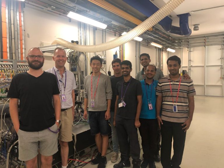
Prof. Peter Lee, who leads the MAPP in-situ synchrotron imaging activities, applied some of these to help clinicians and biologists better understand the damage COVID-19 can do to our organs.
In March 2020 two pathologists in Germany from a group, the UCL group had worked with in the past contacted Prof. Lee about imaging biopsies in 3D, working with Dr Disney this was done at Diamond Light Source.
At the same time Prof. Lee contacted Dr Paul Tafforeau, the European Synchrotron Radiation Facility (ESRF), to use the capabilities of the Extremely Brilliant Source (EBS) upgrade of ESRF to the world’s first 4th generation high energy source.
Prof. Lee formed a team of imaging scientists (Walsh at UCL) and medical specialists (Jonigk and Ackermann in Germany) to develop a technique that they call Hierarchical Phase-Contrast Tomography (HiP-CT) that enabled intact human organs to be scanned with 20-micron voxels everywhere, zooming to 1 micron locally without taking biopsies.
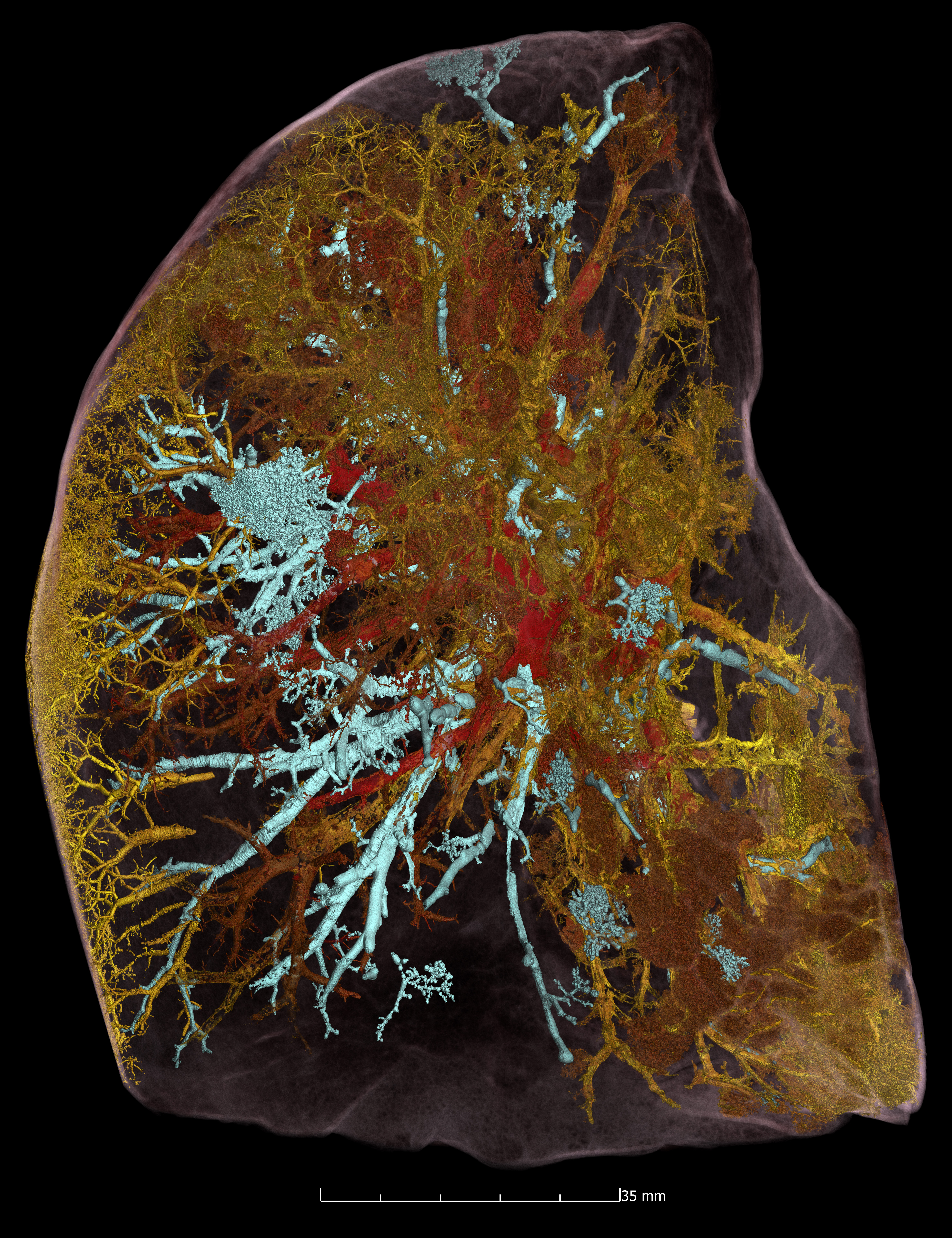
The application of this technique to image the damage to a 54-year-old male COVID-19 victim’s lungs is shown in the image.
Working with the Fraunhofer Inst. in Germany and UK teams, a new beamline, BM18, is being developed to apply it to industrial applications from batteries to aero-engines.
More:
Demonstrators
-
Video/ Animation illustrating Directed Energy Deposition (DED) -
Solid-State Processing of Surplus Aluminium Alloy Powders through a Combination of Field Assisted Sintering Technology and Hot Rolling
The Sheffield Titanium Alloy Research [STAR] group partners with ECKART GmbH to convert surplus aluminium alloy powders from atomisation...
-